
FIGURE
Fig. 4
- ID
- ZDB-FIG-190827-27
- Publication
- Monma et al., 2019 - Aging-associated microstructural deterioration of vertebra in zebrafish
- Other Figures
- All Figure Page
- Back to All Figure Page
Fig. 4
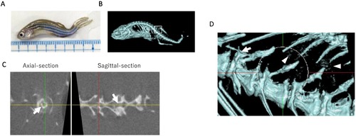
Micro-CT images of 45 mpf zebrafish. A–B. Whole bright field (A) and micro-CT (B) images of aged zebrafish; C. Axial and sagittal-sectional image of FCV, Arrows indicate ossification inside FCV; D. Magnified image of B (white circle is FCV), Arrowheads indicate ectopic ossifications around vertebrae. |
Expression Data
Expression Detail
Antibody Labeling
Phenotype Data
Phenotype Detail
Acknowledgments
This image is the copyrighted work of the attributed author or publisher, and
ZFIN has permission only to display this image to its users.
Additional permissions should be obtained from the applicable author or publisher of the image.
Full text @ Bone Rep