
- Title
-
Aging-associated microstructural deterioration of vertebra in zebrafish
- Authors
- Monma, Y., Shimada, Y., Nakayama, H., Zang, L., Nishimura, N., Tanaka, T.
- Source
- Full text @ Bone Rep
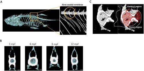
Morphological images with micro-CT visualisation. A. The position of FCV. FCV exhibits elongated unfused haemal arches with shortened ribs, and absence of a haemal spine (Bird and Mabee, 2003). We selected this vertebra in the following analyses. B. Cross-sectional images of FCV in 3–12 mpf zebrafish. Surrounding bone over the threshold of 300 mg/cm3 in BMD is defined as the cortical bone (white area), and the region surrounded by cortical bone is defined as ‘tissue’ (blue area), containing trabecular bone. C. Representative 3-D images of FCV. |
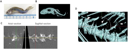
Micro-CT images of 45 mpf zebrafish. A–B. Whole bright field (A) and micro-CT (B) images of aged zebrafish; C. Axial and sagittal-sectional image of FCV, Arrows indicate ossification inside FCV; D. Magnified image of B (white circle is FCV), Arrowheads indicate ectopic ossifications around vertebrae. |

. Sagittal images of vertebrae in 3 – 12 mpf female zebrafish. |