FIGURE

Figure 2

ID
ZDB-FIG-190723-884
Publication
Zhang et al., 2019 - Endoglin is a conserved regulator of vasculogenesis in zebrafish-Implications for hereditary haemorrhagic telangiectasia
Other Figures
All Figure Page
Back to All Figure Page
Figure 2

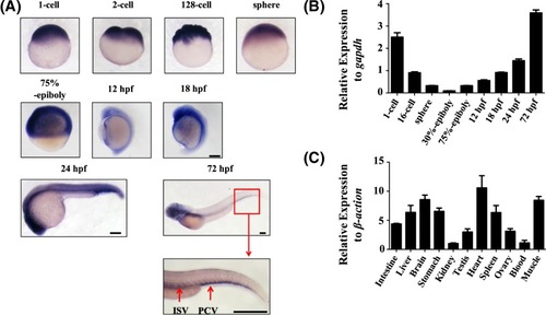
Temporal-spatial expression of endoglin

(A) Embryonic zebrafish endoglin expression by WISH (n>30 for each group). Embryo stages, as labelled, are shown in lateral views from the 1 cell to 72 hpf stage. Blue staining shows a positive signal for endoglin expression and the red box indicates high expression of endoglin in the tail region. Scale bars are 200 μm. (B) qPCR analysis of endoglin expression in different stages of zebrafish embryos from the 1-cell to 72 hpf stage. Gapdh was used as an internal control (mean ± SD; the experiments were repeated three times). (C) The mRNA level of endoglin in various tissues of the adult zebrafish, including the intestine, liver, brain, stomach, kidney, testis, heart, spleen, ovary, blood and muscle. β-Actin was used as an internal control (mean ± SD; the experiments were repeated three times).

Expression Data
Gene:
Fish:
Anatomical Terms:
Stage Range: 1-cell to Adult

Expression Detail
Antibody Labeling
Phenotype Data

Phenotype Detail
Acknowledgments
This image is the copyrighted work of the attributed author or publisher, and ZFIN has permission only to display this image to its users. Additional permissions should be obtained from the applicable author or publisher of the image. Full text @ Biosci. Rep.