|
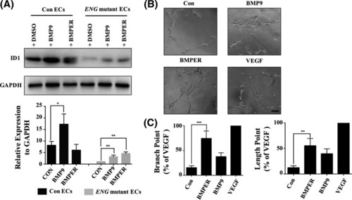
Enhancing the expression of BMPER increased the expression of ID1 and blood vessel formation in ENG mutant ECs (A) Western blot analysis of ID1 expression in the Con ECs + 0.1% DMSO, Con ECs + 10 ng/ml BMP9, Con ECs + 20 ng/ml BMPER, ENG mutant ECs + 0.1% DMSO, ENG mutant ECs + 10 ng/ml BMP9 and ENG mutant ECs + 20 ng/ml BMPER group. Each group was incubated for more than 24 h at 37°C. GAPDH was used as an internal control. Grey scanning analysis, conducted using ImageJ, was used to analyse the Western blot results (mean ± SD; the experiments were repeated three times). (B) The tube formation of ENG mutant ECs in four groups, including the Con (EC(FULL)-SFM medium without 30 ng/ml VEGF), VEGF (EC(FULL)-SFM medium), BMP9 (EC(FULL)-SFM medium without 30 ng/ml VEGF + 10 ng/mL BMP9) and BMPER (EC(FULL)-SFM medium without 30 ng/ml VEGF + 20 ng/ml BMPER) group. Tube formation was assessed and photographed at 3 h. Scale bars are 100 μm. (C) Quantitative results of tube formation. The number and the length of branches were assessed and counted by Imagine J. The VEGF group was a positive control. The ratio of tube formation = (the number/full-length of branches in each group):(the number/ full-length of branches in VEGF group). Error bars represent the SD of the mean values from three independent experiments. A value of P was considered statistically significant (*P<0.05, **P<0.01, ***P<0.001) for A and C.
|