|
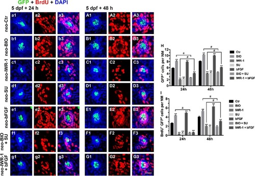
Effects of exogenous regulation of the Wnt and FGF pathways on HC regeneration at 24 hpa and 48 hpa after neomycin injury.a1–G3: Regenerating cells labeled with BrdU (red) and HCs labeled with GFP (green). a1–a3: 24 hpa after neomycin damage; A1–A3: 48 hpa after neomycin damage; b1–b3: 24 hpa after addition of BIO; B1–B3: 48 hpa after addition of BIO; c1–c3: 24 hpa after addition of IWR-1; C1–C3: 48 hpa after addition of IWR-1; d1–d3: 24 hpa after addition of SU5402; D1–D3: 48 hpa after addition of SU5402; e1–e3: 24 hpa after addition of bFGF; E1–E3: 48 hpa after addition of bFGF; f1–f3: 24 hpa after addition of BIO+SU5402; F1–F3: 48 hpa after addition of BIO + SU5402; g1–g3: 24 hpa after addition of IWR-1 + bFGF; G1–G3: 48 hpa after addition of IWR-1 + bFGF. h and i: Quantification of the HCs (GFP+) (h) and regenerating HCs (BrdU+ GFP+) (i) after blocking or activating the Wnt and FGF pathways. n = 7 fish per group. ** Indicates p < 0.001, # indicates p < 0.05, and error bars indicate the standard error of the mean. Scale bar in G3 = 20 μm for a1–G3
|