|
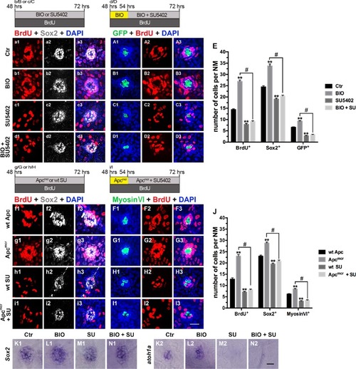
Effects of the Wnt and FGF signaling pathways on proliferation and differentiation during initial neuromast development.a1–D3: Proliferative cells labeled with BrdU (red), SCs labeled with Sox2 (white), and HCs labeled with GFP (green). a1–A3: Control neuromast; b1–B3: After addition of BIO; c1–C3: After addition of SU5402; d1–D3: After 6 h of incubation with BIO, SU5402 and BIO were added for 18 h; e: Quantification of the proliferative cells (BrdU+), SCs (Sox2+), and HCs (GFP+) shown in a1–D3. f1–I3: Proliferative cells labeled with BrdU (red), SCs labeled with Sox2 (white), and HCs labeled with myosin VI (green). f1–F3: wt Apc larvae; g1–G3: Apcmcr mutants; h1–H3: After addition of SU5402; i1–I3: 54 hpf Apcmcr embryos were treated with SU5402 for 18 h. j: Quantification of the proliferative cells (BrdU+), SCs (Sox2+), and HCs (Myosin VI+) shown in f1–I3. n = 5–7 fish per group. ** Indicates p < 0.001, # indicates p < 0.05, and error bars indicate the standard error of the mean. K1–N1 and K2–N2: WISH analysis of Sox2 (K1–N1) and atoh1a (K2–N2) expression in the neuromasts from different groups. Scale bar in I3 = 20 μm for a1–D3 and f1–I3. Scale bar in N2 = 30 μm for K1–N1, K2–N2
|