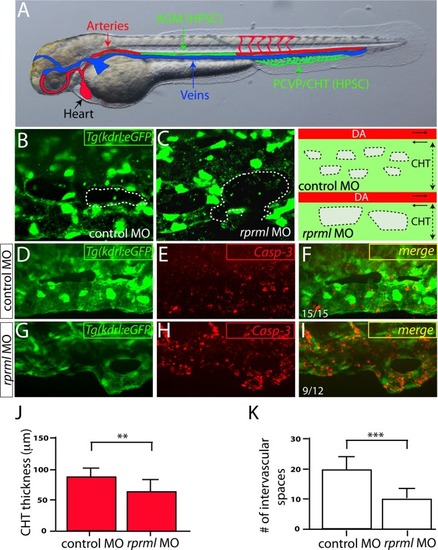
Lack of rprml increases cell apoptosis within the HSPC niche at the CHT. (A) Schematic diagram showing zebrafish vasculature at 48 hpf. In green: (1) aorta-gonad-mesonephros (AGM) where the HSPCs are born, and (2) the posterior caudal vein plexus/caudal hematopoietic tissue (PCVP/CHT), which constitutes the embryonic HSPC niche. Red and blue colors correspond to the artery and vein, respectively. (B–I) Confocal spinning-disk images of the CHT of Tg(kdrl:eGFP) at 48 hpf. (B,C) Note that the spaces are slightly larger in rprml morphants compared to control embryos. Right panel shows a schematic representation of the observed phenotypes from (B,C). Dotted areas indicate intervascular spaces within the CHT. (E,F,H,I) Anti-activated caspase-3 (Casp-3, red) labels apoptotic cells. (D,F) A control MO-injected embryo showing (D) normal morphology of the CHT and (E,F) standard levels of Casp-3 activity. (G–I) rprml-MO injected embryos show (H,I) increased Casp-3 immunoreactivity within the PCVP/CHT. Number of embryos with the phonotype shown as a fraction of the total number of embryos examined is indicated in the bottom left corner in (F,I). (J,K) Statistical significance was determined using two-tailed unpaired Student’s t-test. **P ≤ 0.01, ***P ≤ 0.001.
|
