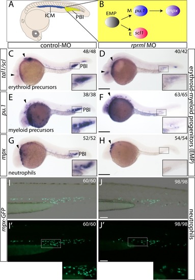
rprml is required for transient-definitive hematopoiesis. (A) Schematic diagram for zebrafish hematopoiesis at 28 hpf showing the intermediate cell mass (ICM) and the posterior blood island (PBI). (B) Schematic for the transient-definitive wave of hematopoiesis where EMP give rise to erythroid myeloid and granulocytic/neutrophils cell population at the PBI. (C,E,G,I) Lateral views of control MO and (D,F,H,J) rprml MO-injected embryos. (C–H) 28 hpf embryos analyzed by WISH against erythroid/myeloid progenitor and granulocyte markers: (C–D) tal1/scl1 for erythroid precursor cells, (E,F) pu.1 for myeloid precursor cell, (G,H) mpx for neutrophils. Inset magnifications from (C) to (H) show the posterior blood island (PBI). Black arrowheads in (D,F,H) show normal expression of tal1/scl1, pu.1 and mpx in the anterior primitive hematopoietic territories. (I,J) Representative images of 48 hpf transgenic Tg(mpx:GFP) embryos analyzed by fluorescent microscopy. (I,I′) control and (J,J′) rprml morphant embryos. Inset magnifications in (I′,J′) show positive fluorescence in neutrophils at the caudal hematopoietic tissue. Number of embryos with the phenotype shown as a fraction of the total number of embryos examined is indicated in the top right corner in (C–J). Scale bars, 200 μm (C–H) and 100 μm (I,J).
|
