|
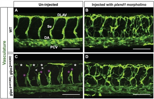
Removal of <italic>plxnd1</italic> activity from <italic>gipc1<sup>skt1(MZ)</sup>; gipc2<sup>skt4(MZ)</sup></italic> maternal-zygotic (MZ) double mutants yields a phenotype similar to that of <italic>plxnd1</italic> nulls.(A–D) Confocal lateral images of the trunk vasculature (green) of 32 hpf embryos (region dorsal to the yolk extension). Anterior, left; dorsal, up. Scale bars (white horizontal lines), 100 μm. Morpholino injection (un-injected or injected with plxnd1 morpholino) indicated on top, genotypes (WT or gipc1skt1(MZ); gipc2skt4(MZ)) indicated on the left. The un-injected WT picture (A) shows the names of the major vessels in white font: DLAV (Dorsal Longitudinal Anastomotic Vessel), Se (Segmental Vessel), DA (Dorsal Aorta), and PCV (Posterior Cardinal Vein). Vascular defects highlighted as follows: truncated or missing Se (magenta asterisk), thin Se (white greater/less-than signs), DLAV gaps (white asterisk). Quantifications. The following number of embryos were analyzed: WT (four embryos), WT injected with plxnd1 morpholino (four embryos; 4/4 showed a vascular phenotype similar to that of plxnd1fov01b nulls), gipc1skt1(MZ); gipc2skt4(MZ) (12 embryos; 7/12 showed angiogenesis deficits), and gipc1skt1(MZ); gipc2skt4(MZ) injected with plxnd1 morpholino (11 embryos; 11/11 showed a vascular phenotype similar to that of plxnd1fov01b nulls).
|