
FIGURE
Fig. 6
- ID
- ZDB-FIG-190718-15
- Publication
- Eno et al., 2019 - Aggregation, segregation and dispersal of homotypic germ plasm RNPs in the early zebrafish embryo
- Other Figures
- All Figure Page
- Back to All Figure Page
Fig. 6
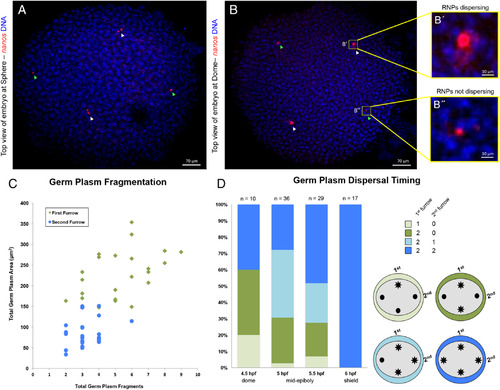
Germ plasm aggregate dispersal correlates with timing of RNP furrow recruitment. A,B: Top view of embryos demonstrating asynchrony in fragmentation and dispersal timing; germ plasm labeled with in situ hybridization for nanos and DNA with DAPI. A:Embryo at sphere stage with germ plasm aggregates localized to regions correlating to furrow origin. Germ plasm aggregates with inferred first furrow origin underwent fragmentation whereas second furrow aggregates did not. B: Embryo at dome stage with germ plasm aggregates correlating to furrow origin. In A,B, aggregates inferred to be derived from first and second furrow are labeled with white and green arrowheads, respectively. Germ plasm aggregate with inferred first furrow origin (B′) is undergoing RNP dispersal, while an aggregate with inferred second furrow origin (B″) is not. C:Correlation between germ plasm inferred furrow origin and aggregate fragmentation, n = 24 embryos. Total germ plasm area correlates (r = 0.66) with the number of germ plasm fragments in the same embryonic quadrant, with larger masses, inferred to have formed in the furrow for the first cell cycle, undergoing more fragmentation. D:Increase in frequency of RNP cytoplasmic dispersal through development and correlation between germ plasm inferred furrow origin and RNP dispersal. Embryos show patterns of germ plasm RNP cytoplasmic dispersal consistent with larger aggregates formed during first cell cycle furrow undergoing dispersal earlier than those formed during the second cell cycle. Other possible patterns of germ plasm RNP dispersal, such as a second furrow aggregate dispersing before either first furrow aggregate, were not observed. See text for details. All images are confocal Z‐projections. Scale bars = 70 μm A & B, 10 μm B′ & B″. |
Expression Data
Expression Detail
Antibody Labeling
Phenotype Data
Phenotype Detail
Acknowledgments
This image is the copyrighted work of the attributed author or publisher, and
ZFIN has permission only to display this image to its users.
Additional permissions should be obtained from the applicable author or publisher of the image.
Full text @ Dev. Dyn.