
Fig. 4
- ID
- ZDB-FIG-190718-12
- Publication
- Eno et al., 2019 - Aggregation, segregation and dispersal of homotypic germ plasm RNPs in the early zebrafish embryo
- Other Figures
- All Figure Page
- Back to All Figure Page
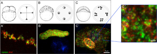
Germ plasm RNPs remain homotypic during asymmetric germ plasm segregation and dispersal. A–C″: The single‐type character of germ plasm RNPs appears to be maintained through aggregate formation at the furrows for the first several cell cycles (0–2 hpf [A,A′]), segregation in the blastula (2–4 hpf [B,B′]), and RNP dispersal at the end of the blastula period [4–6 hpf (C–C″]). Note in panels (C′,C″) RNPs are dispersed, largely occupying the cytoplasmic space, in contrast to aggregated, subcellularly localized germ plasm at earlier stages (A,B; see also Fig. 2 top left inset), Panel A′ = confocal Z‐projection; Panels B′–C″ = SIM Z‐projections. Scale bars = 5 μm in A′–C′; 1 μm in C″. Panels (A–C) adapted from Kimmel et al. (1995), with permission. |