
Fig. 2
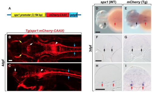
Tg(spx1:mCherry-CAAX) zebrafish express mCherry-CAAX in spx1-expressing cells. (A) Schematic of the plasmid construct for the generation of Tg(spx1:mCherry-CAAX) zebrafish. (B,C) Dorsal (B) and lateral (C) views of Tg(spx1:mCherry-CAAX) zebrafish at 4 days post-fertilisation (dpf), anterior to the left. White arrowheads designate spx1-expressing neurons in the hindbrain, and blue arrows indicate spx1+ axonal projections to the spinal cord. White arrows mark the midbrain tegmentum, and yellow arrows label spx1+ axonal projections to the forebrain. (D–I) In situ RNA hybridization of 3 dpf wildtype and Tg(spx1:mCherry-CAAX) embryos with spx1 (D,F,H) and mcherry mRNA (E,G,I), respectively. Dorsal views of whole-mount embryos with anterior to the left (D,E), and transverse sections of the brain with dorsal to the top (F–I). Black and red arrows indicate labelled cells in the midbrain tegmentum and hindbrain, respectively. Scale bar: 50 μm in B,C,F–I; 100 μm in D,E. |
| Genes: | |
|---|---|
| Fish: | |
| Anatomical Terms: | |
| Stage Range: | Protruding-mouth to Day 4 |