
Fig. 2
- ID
- ZDB-FIG-190711-16
- Publication
- Ghersi et al., 2019 - bif1, a new BMP signaling inhibitor, regulates embryonic hematopoiesis in the zebrafish
- Other Figures
- All Figure Page
- Back to All Figure Page
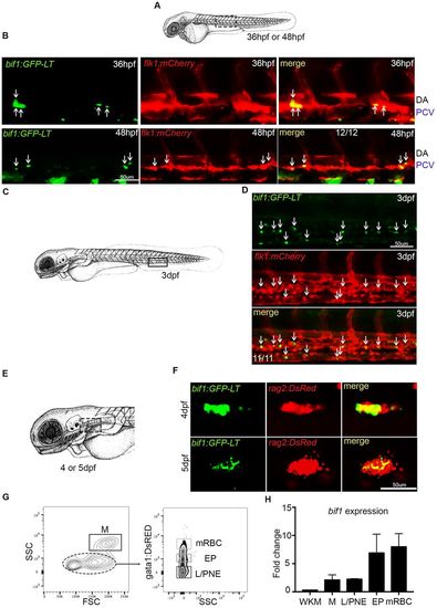
bif1 is also expressed during definitive and adult hematopoiesis. (A) Schematic indicating the approximate area in the trunk shown in the images in B (dotted rectangle). (B) Confocal images of the trunk region of double-positive bif1:GFP-LT and flk1:mcherry embryos at 36 hpf and 48 hpf. Arrows indicate double-positive cells. (C) Schematic indicating the approximate area in the tail shown in the images in D (rectangle). (D) Confocal images of the CHT of double-positive bif1:GFP-LT and flk1:mcherry embryos at 3 dpf. Arrows indicate double-positive cells. (E) Schematic representing the head of a zebrafish at 4 dpf. The position of the thymus is outlined by the dotted rectangle. (F) Confocal images of thymic area of double-positive bif1:GFP-LT;rag2:DsRed embryos at 4 and 5 dpf. (G) Results from FACS-sorted cells of WKM dissected from 1-year-old embryos. n=3. (H) Expression of bif1 by qPCR on FACS-sorted cells, relative to the WKM. Data were obtained from biological triplicates. SSC, side scatter; FSC, forward scatter; M, myeloid; L/PNE, lymphocytes and progenitors non-erythroid; EP, erythroid progenitors; mRBC, mature red blood cells; WKM, whole kidney marrow. |
| Genes: | |
|---|---|
| Fish: | |
| Anatomical Term: | |
| Stage Range: | Prim-25 to Long-pec |