
FIGURE
Fig. 5
- ID
- ZDB-FIG-190708-19
- Publication
- Morales et al., 2019 - Peripheral Macrophages Promote Tissue Regeneration in Zebrafish by Fine-Tuning the Inflammatory Response
- Other Figures
- All Figure Page
- Back to All Figure Page
Fig. 5
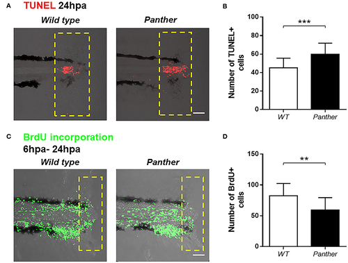
Increased cell death and reduced cell proliferation after tail fin amputation in panther larvae. (A) Cell death was measured at 24 hpa through TUNEL assays. Red dots represent TUNEL+ cells. (B) Quantification of TUNEL+ cells ± SD in the damage site of panther and WT larvae at 24 hpa. Twenty larvae per condition were used. (C) Cell proliferation was assessed by BrdU incorporation from 6 to 24 hpa. Green dots represent BrdU+ cells (D) The number of BrdU+ cells ± SD in panther and WT individuals was obtained from 12 larvae per condition. **p < 0.01; ***p < 0.001. |
Expression Data
Expression Detail
Antibody Labeling
Phenotype Data
Phenotype Detail
Acknowledgments
This image is the copyrighted work of the attributed author or publisher, and
ZFIN has permission only to display this image to its users.
Additional permissions should be obtained from the applicable author or publisher of the image.
Full text @ Front Immunol