
Fig. 2
- ID
- ZDB-FIG-190708-15
- Publication
- Morales et al., 2019 - Peripheral Macrophages Promote Tissue Regeneration in Zebrafish by Fine-Tuning the Inflammatory Response
- Other Figures
- All Figure Page
- Back to All Figure Page
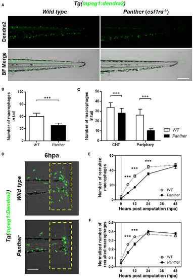
Panther (csf1ra−/−) larvae have a fewer peripheral macrophage population and a delayed recruitment of macrophages after tail fin amputation. (A) Representative images of Tg(mpeg1:Dendra2) larvae in a wild type (WT) or a panther genetic background. Scale bar = 250 μm. (B) Total number of macrophages ± SD in the tail of panther and WT larvae at 72 hpf. (C) Quantification of peripheral tissue-resident and CHT-resident macrophages in the tail of panther and WT larvae. Means ± SDs for each condition are shown in the graph. (D)Recruitment of macrophages (green cells in the yellow dashed rectangle) in panther and WT individuals after tail fin amputation. Scale bar = 100 μm. (E) Quantification of recruited macrophages ± SEM after tail fin amputation in panther and WT larvae from 0 to 48 hpa. Twenty larvae per condition were used. (F) The previous quantification was normalized by the number of total macrophages in the tail of the respective larva (the sum of peripheral, CHT, and recruited macrophages). ***p < 0.001. |