
Fig. 3
- ID
- ZDB-FIG-190708-16
- Publication
- Morales et al., 2019 - Peripheral Macrophages Promote Tissue Regeneration in Zebrafish by Fine-Tuning the Inflammatory Response
- Other Figures
- All Figure Page
- Back to All Figure Page
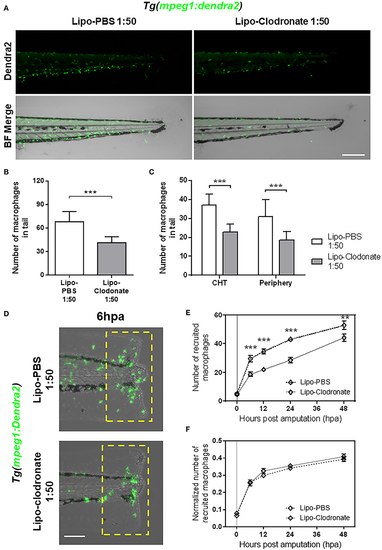
Partial reduction of macrophages pool does not affect the kinetic of macrophage recruitment after tail fin amputation. (A) Images of 72 hpf Tg(mpeg1:Dendra2) larvae, 18 h after injection of an 1:50 dilution of Lipo-PBS and Lipo-clodronate in the bloodstream, respectively. Scale bar = 250 μm. (B) Total macrophages ± SD in the tail of Lipo-clodronate 1:50 and Lipo-PBS 1:50 larvae at 72 hpf. (C) Mean ± SD of peripheral tissue-resident and CHT-resident macrophages in the tail. (D) Recruited macrophages (green cells in the yellow dashed rectangle) in Lipo-clodronate 1:50 and Lipo-PBS 1:50 individuals after tail fin amputation. Scale bar = 100 μm. (E) Quantification of recruited macrophages ± SEM after tail fin amputation in Lipo-clodronate 1:50 and Lipo-PBS 1:50 larvae from 0 to 48 hpa. A total of 20 larvae per condition were used. (F) Normalized number of recruited macrophages at each time point. **p < 0.01; ***p < 0.001. |