
Fig. 4
- ID
- ZDB-FIG-190702-25
- Publication
- Umali et al., 2019 - Loss of foxc1 in zebrafish reduces optic nerve size and cell number in the ganglion cell layer
- Other Figures
- All Figure Page
- Back to All Figure Page
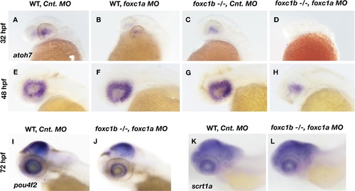
Loss of foxc1 affects markers of RGC differentiation.Expression of atoh7 is induced in the nasal-ventral domain of the retina at 32 hpf (A), and encompasses the entire retina by 48 hpf (E) in wildtype embryos injected with a control morpholino. Loss of foxc1a on its own through morpholino inhibition (B and F) had no effect on atoh7 initiation at 32 hpf, nor its expression throughout the retina at 48 hpf. Similarly, no change in atoh7 expression is observed in foxc1 −/− injected with control morpholinos at either 32 hpf (C) or 48 hpf (G). Injection of foxc1a morpholinos into a foxc1b −/− background impaired the initiation of atoh7 expression at 32 hpf (D), and reduced expression throughout the retina at 48 hpf (H). pou4f2is expressed throughout the RGC layer at 3 dpf (I), and is reduced in foxc1b -/- injected with foxc1a morpholinos (J). A marker of Amacrine cells, scrta1, is not affected (K and L). |
| Genes: | |
|---|---|
| Fish: | |
| Knockdown Reagent: | |
| Anatomical Terms: | |
| Stage Range: | Prim-15 to Protruding-mouth |
| Fish: | |
|---|---|
| Knockdown Reagent: | |
| Observed In: | |
| Stage Range: | Prim-15 to Protruding-mouth |
Reprinted from Vision Research, 156, Umali, J., Hawkey-Noble, A., French, C.R., Loss of foxc1 in zebrafish reduces optic nerve size and cell number in the ganglion cell layer, 66-72, Copyright (2019) with permission from Elsevier. Full text @ Vision Res.