
FIGURE
Fig. 9
- ID
- ZDB-FIG-190701-18
- Publication
- Liu et al., 2019 - Imaging neural events in zebrafish larvae with linear structured illumination light sheet fluorescence microscopy
- Other Figures
- All Figure Page
- Back to All Figure Page
Fig. 9
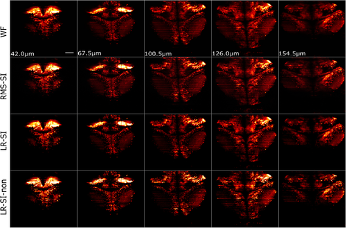
Slices from a 3-D image stack of a 7-day-old elavl3:GCaMP5g; gad1b:RFP; mitfaw2/w2 zebrafish larva with DSLM-SI. From the top to bottom row: WF, RMS-SI, LR-SI, and LR-SI-non. Each column is a different depth into the sample. image size is 399.36 μm×399.36 μm and the scale bar is 50 μm.
|
Expression Data
Expression Detail
Antibody Labeling
Phenotype Data
Phenotype Detail
Acknowledgments
This image is the copyrighted work of the attributed author or publisher, and
ZFIN has permission only to display this image to its users.
Additional permissions should be obtained from the applicable author or publisher of the image.
Full text @ Neurophotonics