
Fig. 1
- ID
- ZDB-FIG-190624-3
- Publication
- Huang et al., 2019 - Rab33a and Rab33ba mediate the outgrowth of forebrain commissural axons in the zebrafish brain
- Other Figures
- All Figure Page
- Back to All Figure Page
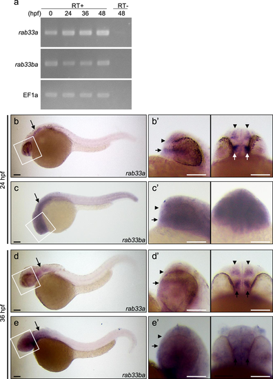
Expression of zebrafish rab33a and rab33ba. (a) RT-PCR analysis of rab33a and rab33ba transcripts. Elongation factor 1a (EF1a) was used as a control. Developmental stages are denoted as hours post-fertilization (hpf). RT-PCR products produced in the presence (RT+) or absence (RT−) of reverse transcriptase were electrophoresed on 2% agarose gels. (b,c) Whole-mount in situ hybridization of rab33a (b) and rab33ba (c) at 24 hpf; (b′) and (c′) show the enlarged lateral (left) and ventral (right) views of the areas indicated by the rectangles. (d,e) Whole-mount in situ hybridization of rab33a (d) and rab33ba(e) at 36 hpf; (d′) and (e′) show the enlarged lateral (left) and ventral (right) views of the areas indicated by the rectangles. Arrowheads and arrows in (b′–e′) indicate the DRC and VRC, respectively. Arrows in (b–e) indicate the hindbrain. See the negative control data for whole-mount in situ hybridization (Supplementary Fig. S3). Scale bars: 100 μm.
|
| Genes: | |
|---|---|
| Fish: | |
| Anatomical Terms: | |
| Stage Range: | 1-cell to Long-pec |