
Fig. 2
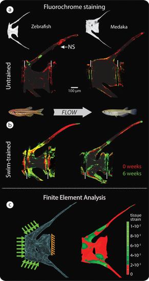
Vertebral bone formation in response to load. (a–b) Bone formation detected by sequential intraperitoneal fluorochrome injections in untrained (a) and swim-trained (b) zebrafish (left, osteocytic) and medaka (right, anosteocytic), each of the 4 groups comprising n = 4 fish. Alizarin red was injected at t = 0 weeks, and calcein green was injected at t = 6 weeks of swim training. Dashed, vertical white lines mark the border between vertebrae. (c) FE model (left) and FEA results (right), showing von Mises strains in a loaded medaka vertebra. Note similarity of peak strains predicted by FEA in (c) and regions of intense bone formation in response to load, indicated by fluorochrome double labeling (b). The distal HSs are cropped in fluorochrome and FE renderings. FE, finite element; FEA, finite element analysis; HS, hemal spine; NS, neural spine. |