
Fig. 4
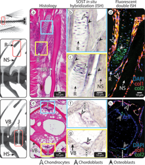
SOST expression in medaka vertebra and dorsal fin R. (a–d) An NS of medaka vertebra and adjacent fin R. (a) HE-stained section. The blue and yellow squares mark 2 regions in the dorsal fin R, shown in (b) and (c), respectively. (b–c) ISH showing SOST expression in the cartilaginous core of the dorsal fin R (white arrowheads) and surface osteoblasts of the fin R and NS (black arrowheads). (d) Double fluorescent ISH, showing both Col1a1 (red) and Col2a1 (green) expression in the SOST-positive osteoblasts (black arrowheads) of the NS and fin R and Col2a1 in the SOST-positive chondrocytes (white arrowhead) in the cartilaginous core of the fin R. (e–h) The IVR in the caudal vertebral column of medaka, (e) HE-stained section. The blue and yellow squares mark the 2 regions of the intravertebral region shown in (f) and (g), respectively. (f–g) ISH showing SOST expression in chordoblasts (gray arrowheads), identified by their distinct morphology and location. (h) Double fluorescent ISH in the IVR, showing Col1a1 expression (red) in osteoblasts (black arrowhead) in the external region of the IVR and Col2a1 expression (green) in SOST-positive chordoblasts (gray arrowhead) in the internal region of the IVR (scale bar same as in e). Dashed, vertical white lines mark the border between vertebrae. Results for individual stains (e.g., DAPI, SOST, col1, col2) are shown in S4 Fig. Col1a1, collagen type I alpha 1; Col2a1, collagen type II alpha 1; HE, hematoxylin–eosin; ISH, in situ hybridization; IVR, intervertebral region; J, joint; NS, neural spine; R, radial; VB, vertebral bone |