
Fig. 2
- ID
- ZDB-FIG-190604-2
- Publication
- Fei et al., 2018 - A cargo model of yolk syncytial nuclear migration during zebrafish epiboly
- Other Figures
- All Figure Page
- Back to All Figure Page
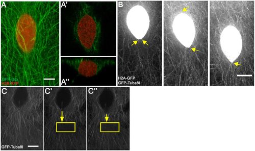
E-YSN move along and beneath the microtubule network. (A) Confocal projection of e-YSN labeled with H2B-RFP and microtubules labeled with GFP-Dcx. (A′) Deep single plane shows microtubules around the nucleus. (A″) Orthogonal view shows e-YSN is largely below the microtubule network. (B) Stills from Movie 2 of Tg:(XlEefla1:GFP-tuba81) embryo injected with h2a-gfp RNA. The e-YSN becomes elongated and leading tip of e-YSN becomes pointed during migration (yellow arrows). (C-C″) Selected confocal projections of a photobleached Tg:(XlEefla1:GFP-tuba81) embryo injected with h2a-gfp RNA. Yellow rectangle marks the photobleached region and shortening yellow arrow shows that the e-YSN moves towards the photobleached region. Pre-bleach, C; immediately post-bleach, C′; 30 s post-bleach, C″. Scale bars: 7 µm in A; 10 µm in B; 9 µm in C. |