
FIGURE
Fig. 5
- ID
- ZDB-FIG-190429-6
- Publication
- Chen et al., 2018 - Folic acid-nanoscale gadolinium-porphyrin metal-organic frameworks: fluorescence and magnetic resonance dual-modality imaging and photodynamic therapy in hepatocellular carcinoma
- Other Figures
- All Figure Page
- Back to All Figure Page
Fig. 5
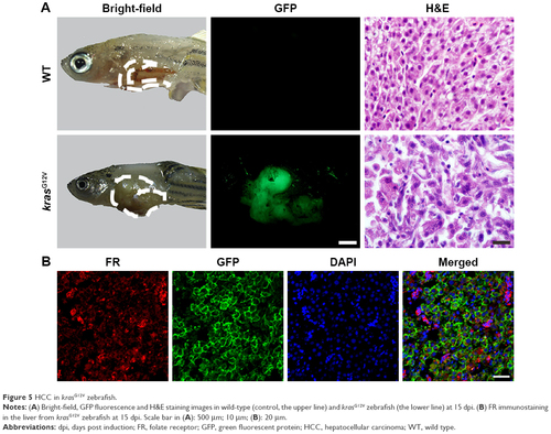
HCC in krasG12V zebrafish. Notes: (A) Bright-field, GFP fluorescence and H&E staining images in wild-type (control, the upper line) and krasG12V zebrafish (the lower line) at 15 dpi. (B) FR immunostaining in the liver from krasG12V zebrafish at 15 dpi. Scale bar in (A): 500 μm; 10 μm; (B): 20 μm. Abbreviations: dpi, days post induction; FR, folate receptor; GFP, green fluorescent protein; HCC, hepatocellular carcinoma; WT, wild type. |
Expression Data
Expression Detail
Antibody Labeling
Phenotype Data
Phenotype Detail
Acknowledgments
This image is the copyrighted work of the attributed author or publisher, and
ZFIN has permission only to display this image to its users.
Additional permissions should be obtained from the applicable author or publisher of the image.
Full text @ Int. J. Nanomedicine