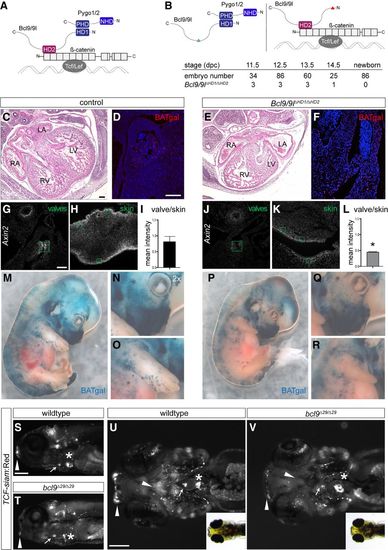
The BCL9–Pygo complex drives Wnt/β-catenin transcription during heart development. (A) Schematic representation of the β-catenin transcriptional complex to outline the experimental strategy. BCL9/9l simultaneously interacts with Pygo1/2 and β-catenin via the HD1 and HD2, respectively. (B) In Bcl9/9l-Δ1/Δ2 mice, no Bcl9/9l molecule can simultaneously bind Pygo1/2 and β-catenin; the deletion of the HD2 or HD1 abrogates Bcl9/9l binding to β-catenin (left panel) or Pygo1/2 (right panel), respectively. Bcl9/9l-Δ1/Δ2 embryos are never found after 14.5 dpc (table). (C–F) Hematoxylin/eosin staining on heart sections of control (C) and Bcl9/9l-Δ1/Δ2 (E) embryos at 13.5 dpc. Bcl9/9l-Δ1/Δ2 embryos have pronounced heart defects, with thinned myocardium of the ventricular walls and malformations of the forming septum, the atrio–ventricular valves, and OFT. The expression of in vivo BATgal Wnt reporter in the OFT region is markedly reduced in Bcl9/9l-Δ1/Δ2 mutant (F) compared with control (D) hearts. (RA) Right atrium; (LA) left atrium; (RV) right ventricle; (LV) left ventricle. (G–L) Axin2 expression (detected by single-molecule mRNA ISH) in wild-type and Bcl9/9l-Δ1/Δ2 OFT valves (G,J) and skin (H,K) at 13.5 dpc. Green boxes are drawn around transversal sections of individual valves of the OFT (G,J) and in skin sections from the limbs (H,K) for quantification of fluorescence intensity levels (I,L). Axin2 expression is strongly reduced in the valves of Bcl9/9l-Δ1/Δ2 embryos (J), while reduction in the skin is mild (K). (I,L) The quantification of the ratio between Axin2 expression in the valves and the skin in the different genotypes reveals a significant reduction of Axin2 expression in Bcl9/9l-Δ1/Δ2 valves. (*) P-value < 0.05. (M–R) In vivo BATgal reporter expression in 13.5-dpc wild-type (M–O) and Bcl9/9l-Δ1/Δ2 mutant (P–R) embryos. Mutant embryos have a slightly decreased reporter expression, but BATgal reporter expression is generally retained in most tissues. Reduced BATgal activity is observed in the craniofacial region (N,Q) and the forelimbs (O,R). Loss of canonical Wnt signaling transcription in the forelimbs is accompanied by severe developmental limb defects in Bcl9/9l-Δ1/Δ2 embryos (cf. O and R). (S–V) Fluorescent and bright-field images of TCF-siam:Red bcl9Δ29 and wild-type zebrafish embryo lateral (S,T) and ventral (U,V) views; anterior is to the left. TCF reporter activity is specifically reduced in the cardiac OFT (arrow) and craniofacial apparatus (arrowheads) and altered in the atrio–ventricular valve (asterisks). Bars: C–F,G,H,J,K, 100 µm; S–V, 200 µm.
|
