
Fig. S5
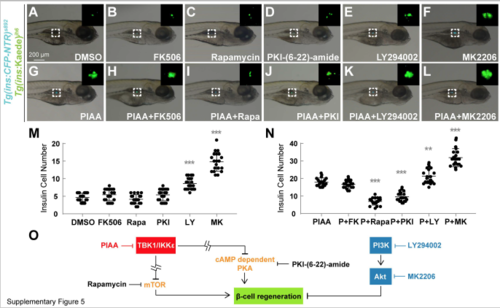
TBK1/IKKε suppression promotes b-cell regeneration via PKA and mTOR signaling axis. (A-L) Bright-field images combined with fluorescent images showing the overall morphology and [Tg(ins:CFP-NTR)s892; Tg(ins:Kaede)jh6] expression (green) of larvae at 48 hpa treated with DMSO (A), calcineurin inhibitor FK506 (B), mTOR inhibitor rapamycin (C), PKA inhibitor PKI-(6-22)-amide (D), PI3K inhibitor LY294002 (E), Akt inhibitor MK2206 (F), PIAA (G), and combinations of each inhibitor with PIAA (H-L), respectively. Treatment of rapamycin and PKI-(6-22)-amide suppressed PIAA-mediated b-cell regeneration (G vs. I and J). The individual LY294002 and MK2206 caused increase in the [Tg(ins:CFP-NTR)s892; Tg(ins:Kaede)jh6]-expressing cell population during regeneration (E and F). The combinations of LY294002 or MK2206, especially MK2206, with PIAA resulted in significant increase in b-cell regeneration (K and L) compared to individual treatments (E and F). The insets display magnified views of the pancreatic islets (outlined by the dashed squares). (M-N) Quantification of the number (mean±SD) of total regenerated b-cells at 48 hpa (in A-L; 4.7±1.2 (DMSO), 5.5±1.7 (FK506), 4.2±1.3 (rapamycin), 5.4±1.7 (PKI-(6-22)-amide), 8.7±1.7 (LY294002), 14.8±2.7 (MK2206), 17.9±2.4 (PIAA), 16.7±2.3 (PIAA with FK506), 7.0±2.1 (PIAA with rapamycin), 9.6±2.8 (PIAA with PKI-(6-22)-amide), 21.3±4.3 (PIAA with LY294002), 31.9±5.1 (PIAA with MK2206)). Cells in 20 planes of confocal images from 20 individual larvae were counted per condition. **, P < 0.01; ***, P < 0.001. (O) A model depicting the role of PKA and mTOR signaling pathways in mediating the b-cell regeneration response to TBK1/IKKε inhibition. The site of action of PIAA is shown in red, while those of LY294002 and MK2206 in blue. |