
Fig. S11
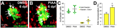
PIAA increases b-cell proliferation during normal development in zebrafish. (A-B) Confocal images of [Tg(ins:CFP-NTR)s892; Tg(ins:Kaede)jh6] larvae at 6 dpf, concurrently treated EdU and DMSO (A) or PIAA (B), respectively, from 4-6 dpf. A small but significant increase in EdU incorporation (white arrows) was observed in PIAA-treated larvae (B) compared to DMSO-treated larvae (A) during normal development. (C) Quantification of the number (mean±SD) of total b-cells (green dots) and b-cells that incorporated EdU (yellow dots) (in A-B; 38.0±3.2 total b-cells, of which 5.8±1.0 (DMSO) and 45.3±9.3, of which 11.0±3.6 (PIAA) incorporated EdU). (D) The percentage (mean±SD) of b-cells that incorporated EdU (in A-B; 15.2±2.7% (DMSO) and 23.8±3.4% (PIAA)). Cells in 20 planes of confocal images from 5 individual larvae were counted per condition. *, P < 0.05. |