
Fig. 3
- ID
- ZDB-FIG-181018-16
- Publication
- Du et al., 2018 - A transgenic zebrafish model for in vivo long-term imaging of retinotectal synaptogenesis
- Other Figures
- All Figure Page
- Back to All Figure Page
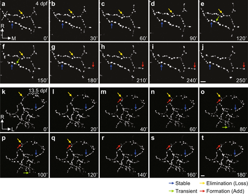
Remodelling of Sypb-EGFP puncta on the axonal arbor of individual RGCs. (a–t) Time-lapse two-photon images of individual RGC axonal arbor expressing Sypb-EGFP showing synaptic remodelling at both early (4 dpf, a–j) and late (13.5 dpf, k–t) larval stages, respectively. At 13.5 dpf, we used pigment mutants of PGUSG (PGUSG; casper). Arbors were imaged at 10 min intervals for at least 3 h. Time in minutes is indicated in the bottom right corner of each panel. The coloured arrows indicate examples of puncta with different behaviours: stable (blue), existing through the entire imaging time period; elimination (yellow), existing at the beginning but lost during imaging; transient (green), newly added during imaging but lost before the end of imaging; formation (red), newly added during imaging and maintained till the end of imaging. L, lateral; M, medial; R, rostral. Scale bar, 5 μm. |