
FIGURE
Fig. 4
- ID
- ZDB-FIG-181012-16
- Publication
- Moore et al., 2018 - Simultaneous ultra-high frequency photoacoustic microscopy and photoacoustic radiometry of zebrafish larvae in vivo.
- Other Figures
- All Figure Page
- Back to All Figure Page
Fig. 4
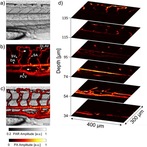
a) PAR image of the trunk of a 5 dpf casper zebrafish. b) PA image of the trunk vasculature acquired simultaneously with a). c) Composite PAR/PA image. d) PA images at different depths within the larva. The scale bar in c) is 50 μm. Ms = Myosepta, M = Myotome, C = Central canal, N = Notochord, Y = Yolk, DLAV = Dorsal longitudinal anastomotic vessel, SV = Intersegmental vein, SA = Intersegmental artery, DA = Dorsal aorta, PCV = Posterior cardinal vein. |
Expression Data
Expression Detail
Antibody Labeling
Phenotype Data
Phenotype Detail
Acknowledgments
This image is the copyrighted work of the attributed author or publisher, and
ZFIN has permission only to display this image to its users.
Additional permissions should be obtained from the applicable author or publisher of the image.
Full text @ Photoacoustics