
Fig. 6
- ID
- ZDB-FIG-180911-46
- Publication
- Oosterhof et al., 2018 - Colony-Stimulating Factor 1 Receptor (CSF1R) Regulates Microglia Density and Distribution, but Not Microglia Differentiation In Vivo
- Other Figures
- All Figure Page
- Back to All Figure Page
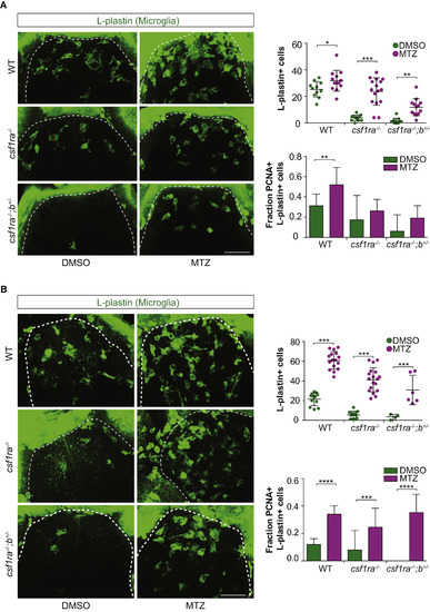
The Response to Neuronal Cell Death of csf1r Mutant Microglia Depends More on Recruitment Than on Proliferation (A and B) We used our previously described conditional neuronal ablation model (van Ham et al., 2012, van Ham et al., 2014), in which treatment with metronidazole (MTZ) leads to selective ablation of neurons with transgenic expression of NTR (the nsfB gene encoding NTR). WT, csf1ra−/−, and csf1ra−/−;b+/− larvae were treated with MTZ at 5 dpf for 16 hr and fixed for immunohistochemistry (whole-mount) at 6 dpf (A) and 7 dpf (B). Immunostaining was performed for dividing (Pcna+) microglia (L-plastin+), and the entire forebrain (dotted lines) was imaged and quantified. Scale bars, 40 μm. Group sizes were at least n = 10 zebrafish larvae (A) and at least n = 4 (B). Error bars indicate SD. ∗p < 0.05, ∗∗p < 0.01, ∗∗∗p < 0.001, ∗p < 0.0001 (one-way ANOVA, Bonferroni multiple testing correction). |
| Antibody: | |
|---|---|
| Fish: | |
| Condition: | |
| Anatomical Term: | |
| Stage Range: | Day 6 to Days 7-13 |
| Fish: | |
|---|---|
| Condition: | |
| Observed In: | |
| Stage: | Day 6 |