
FIGURE
Fig. 9
Fig. 9
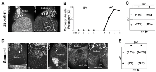
Origin of the coronary vasculature in the blue gourami and zebrafish. Image of a Tg(fli1:EGFP)y1 fish with no identifiable vessel over the ventral aspect of the bulbus (A-i), and the same fish rotated 180 degrees with a visible vessel on the dorsal aspect of the ventricle and connected to the inferior aspect of the atrioventricular junction (A-ii). Frequency of coronary vessel appearance over the bulbus only (BV), and the atrioventricular junction only (AV) over time in the Tg(fli1:EGFP)y1 fish (B). Overall frequency and distribution of coronary vessels in the Tg(fli1:EGFP)y1 between 5 and 8 weeks post fertilization (C). Image of a BS lectin stained gourami heart with a vascular plexus over the surface of the cranial aspect of the bulbus (D-i) and the vessels at higher magnification (D-ii). No vessels are present over the ventricle. The same heart rotated 180 degrees shows the same vessel plexus at the cranial end of the dorsal aspect of bulbus, but no evidence of coronary vessel at or near the atrioventricular junction or the ventricle (D-iii) (B). Further development of the coronary vasculature of the gourami with coronary vessels investing the ventral and dorsal aspect of the ventricle (D-iv,v). Overall frequency and distribution of coronary vessels in the gourami ranging from 40–53 mm in standard length (E).
|
Expression Data
Expression Detail
Antibody Labeling
Phenotype Data
Phenotype Detail
Acknowledgments
This image is the copyrighted work of the attributed author or publisher, and
ZFIN has permission only to display this image to its users.
Additional permissions should be obtained from the applicable author or publisher of the image.
Full text @ J Dev Biol