
- Title
-
Heart Development, Coronary Vascularization and Ventricular Maturation in a Giant Danio (Devario malabaricus).
- Authors
- Shifatu, O., Glasshagel-Chilson, S., Nelson, H.M., Patel, P., Tomamichel, W., Higginbotham, C., Evans, P.K., Lafontant, G.S., Burns, A.R., Lafontant, P.J.
- Source
- Full text @ J Dev Biol
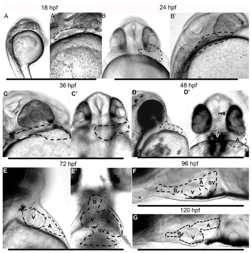
Morphogenesis of the giant danio Devario malabaricus (DM) developing Heart. Representative difference interference contrast (DIC) image of a sagittal view of a newly hatched DM larvae (A). The heart is tubular, laying posterior to the developing eye, extending dorsally, displaying rhythmic and coordinated contraction (Supplemental material movie 1). The tubular heart (outline) at 18 hpf at higher magnification (A’). Representative DIC image of a ventral view of a 24 hpf DM larvae (B) with the tubular heart (outline) projecting to the left. Sagittal view of the same heart laying posterior to eye, extending dorsally (B’). Representative DIC image of a sagittal view of a 36 hpf larvae showing a primarily tubular heart (C,C’). Representative DIC image of a sagittal view of a 48 hpf larvae showing a segmented heart (D). From a ventral view, the ventricle can be seen to have shifted to the right and the atrium remaining on the left (D’), the atrioventricular junction approaching the midline of the anteroposterior (AP) axis. Representative DIC image of a sagittal view of a 72 hpf larvae showing segmented heart (E) with the ventricle occupying a more rostral position. From a ventral view, the ventricle can be seen to have migrated to the right and the atrium remaining on the left ((E’), and Supplemental Material movie 3), with the atrioventricular junction occupying the midline of the AP axis. At 96 hpf (F) and 120 hpf days (G), the heart has assumed its final position, with the atrium is located posterior dorsal to the ventricle, and the bulbus anteriorly (Supplemental Material movie 4). A = atrium, B = bulbus, SV = sinus venosus, V = ventricle.
|

Myosin expression and giant danio DM heart morphogenesis. Myosin (MYH1) immunoreactivity of DM heart at 24 hpf (A), the fish rotated 45 degrees from the AP midline. MYH1 immunoreactivity in a 48 hpf DM larvae (B) showing segmentation of the heart with the ventricle and atrium shifted more toward the midline compared to 24 hpf. MYH1 immunoreactivity in a 72 hpf DM larvae showing pectoral and jaw skeletal muscle, and the segmented heart (ventricle, atrium, and sinus venosus) with the ventricle shifted to the right of the AP midline plane and the atrium remaining on the left (C). Note abundant luminal cardiac trabeculae projecting into the center of the ventricle while they are absent in the atrium and sinus venosus. Myosin staining of DM heart at 2 wpf (D), framed by pectoral and jaw muscles. Only the ventricle can be observed from the ventral view as the atrium is rotated posteriorly. (E) Graphical representation of morphogenesis and anatomical changes to the developing heart in the first 4 dpf based on DIC live imaging and myosin staining. A = atrium, B = bulbus, SV = sinus venosus, sk. mus = skeletal muscle, V = ventricle.
|

Growth of the early DM larvae. Representative images of 1 week post-fertilization (wpf) giant danio (A) and 4 wpf early larvae (B). Change in standard length of giant danio larvae from 36 hpf to 4 wpf (C). Data represent means and standard error from four independent experiments, analyzed using ANOVA, with p < 0.05 considered significant. Scale bar = 5 mm.
|
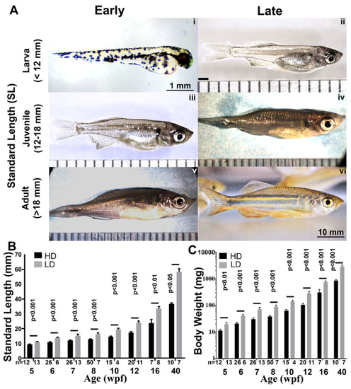
Rearing-density effects on the growth and maturation of the giant danio DM. Images of representative DM early larvae (A-i) and late larvae (A-ii) showing dramatic growth and changes in phenotype. In addition to the abundant melanophores seen in the early larvae, iridophores are prominent in the late larvae as well as the early juvenile (A-iii) giving the fish a distinctly silvery hue. Progressive phenotypic changes, increase in size and the appearance of longitudinal bands of xanthophores characterizes the late juvenile DM (A-iv) and early adult (A-v) and late adult (A-vi). Standard length (SL) measured in low and high density reared fish from 5 to 40 weeks post-fertilization (wpf), and showing differential but linear growth in length (B). Body weight (BW) measured in low and high density reared fish from 5–40 wpf showing differential but logarithmic increase in BW (C). Scale bars are 1 or 10 mm, and ruler units are 1 mm intervals. SL and BW of high and low density raised fish are analyzed using Welch two sample t-test where p < 0.05 is considered significant.
|

Rearing-density effects on the ventricular growth of giant danio DM. Image of an excised adult DM heart (A). The vertical line drawn from the middle of the bulbus at the base of the ventricle to the apex defines the ventricular length (VL). VL of DM measured at high and low density rearing conditions from 5 to 16 wpf (B). VL of high and low density raised fish are analyzed using Welch two sample t-test where p < 0.05 is considered significant. Scattered plot with polynomial fits of DM VL plotted against their standard lengths (SL) showing a positive correlation (C).
|
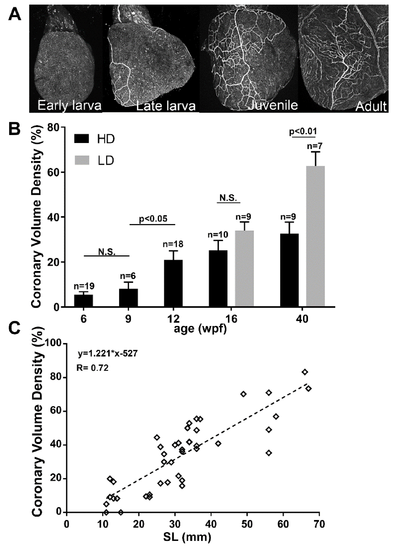
Coronary vascular development in the giant danio DM Heart. Ventral view of a representative B.S. lectin stained heart of an avascular early DM larvae ((A), panel 1), a late larvae with a developing coronary vascular network ((A), panel 2), further expansion of the network in a juvenile ((A), panel 3), and in an adult DM ((A), panel 4). Quantitation of coronary vascularization of the heart from 6–40 wpf, and the effects of rearing density (B). Coronary volume density (CVD) of high and low density raised fish are analyzed using Welch two sample t-test where p < 0.05 is considered significant. Scattered plot of CVD of giant danio DM over standard length (C) showing a positive correlation.
|

Structure of the Adult giant danio DM. Scanning electron micrograph of adult giant danio heart illustrating the ventricle and the dense and highly trabeculated spongy heart, the thin epicardium (Epi) and relatively thick compact heart (Ch). The atrioventricular valves (Va) and part of the atrium (Atr) can also be seen (A). Higher magnification image (B) of the compact heart showing multiple layers of cardiac myocytes forming a thick myocardium, traversed by numerous coronary vessels (Vs), one of which contains a red blood cell (RBC). The junctional space (JS) between the compact and trabeculae is noted. Scale bar = 10 μm. Image of adult giant danio heart section double stained with wheat germ agglutinin (WGA, green) highlighting the borders of compact heart myocytes, and Bandeiraea simplicifolia lectin (BS, red) labeling endothelial cells of the coronary vasculature in high density (C) and low density-reared fish (D). Scale bar = 20 μm. Quantitation of compact heart thickness (E) and minimal coronary vessels diameter (F) in 10-month old giant danio DM. Data analyzed using t-test with p < 0.05 considered significant.
|

Origin of the coronary vasculature in the giant danio DM. Image of a BS lectin stained heart with a single vessel (arrow) over the ventral surface of the bulbus, in contact with the base of the ventricle, and of the same heart rotated 180 degrees, with no evidence of coronary vessel on the dorsal aspect of the ventricle, or near or around atrioventricular junction ((A), left panel). View of a BS lectin stained heart with no identifiable vessel over the bulbus ((B), left panel), and the same heart rotated 180 degrees with vessels connected to the atrioventricular junction on the dorsal aspect of the ventricle ((B), right panel). Image of a BS lectin stained heart with a single vessel (arrow) over the ventral surface of the bulbus, in contact with the base of the ventricle ((C), left panel), and of the same heart rotated 180 degrees, with vessels at the base and dorsal aspect of the ventricle and connected to the atrioventricular junction ((C), right panel). Frequency and distribution of coronary vessels over the bulbus only (BV) and the atrioventricular junction only (AV) from 5 to 16 wpf (D). Frequency and distribution of coronary vessels over the bulbus (BV) and the atrioventricular junction (AV) over standard length (E).
|
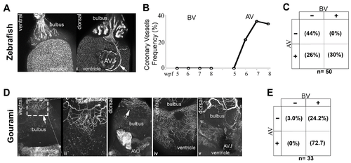
Origin of the coronary vasculature in the blue gourami and zebrafish. Image of a Tg(fli1:EGFP)y1 fish with no identifiable vessel over the ventral aspect of the bulbus (A-i), and the same fish rotated 180 degrees with a visible vessel on the dorsal aspect of the ventricle and connected to the inferior aspect of the atrioventricular junction (A-ii). Frequency of coronary vessel appearance over the bulbus only (BV), and the atrioventricular junction only (AV) over time in the Tg(fli1:EGFP)y1 fish (B). Overall frequency and distribution of coronary vessels in the Tg(fli1:EGFP)y1 between 5 and 8 weeks post fertilization (C). Image of a BS lectin stained gourami heart with a vascular plexus over the surface of the cranial aspect of the bulbus (D-i) and the vessels at higher magnification (D-ii). No vessels are present over the ventricle. The same heart rotated 180 degrees shows the same vessel plexus at the cranial end of the dorsal aspect of bulbus, but no evidence of coronary vessel at or near the atrioventricular junction or the ventricle (D-iii) (B). Further development of the coronary vasculature of the gourami with coronary vessels investing the ventral and dorsal aspect of the ventricle (D-iv,v). Overall frequency and distribution of coronary vessels in the gourami ranging from 40–53 mm in standard length (E).
|