
FIGURE
Fig. 1
Fig. 1
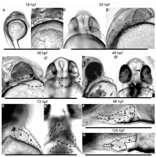
Morphogenesis of the giant danio Devario malabaricus (DM) developing Heart. Representative difference interference contrast (DIC) image of a sagittal view of a newly hatched DM larvae (A). The heart is tubular, laying posterior to the developing eye, extending dorsally, displaying rhythmic and coordinated contraction (Supplemental material movie 1). The tubular heart (outline) at 18 hpf at higher magnification (A’). Representative DIC image of a ventral view of a 24 hpf DM larvae (B) with the tubular heart (outline) projecting to the left. Sagittal view of the same heart laying posterior to eye, extending dorsally (B’). Representative DIC image of a sagittal view of a 36 hpf larvae showing a primarily tubular heart (C,C’). Representative DIC image of a sagittal view of a 48 hpf larvae showing a segmented heart (D). From a ventral view, the ventricle can be seen to have shifted to the right and the atrium remaining on the left (D’), the atrioventricular junction approaching the midline of the anteroposterior (AP) axis. Representative DIC image of a sagittal view of a 72 hpf larvae showing segmented heart (E) with the ventricle occupying a more rostral position. From a ventral view, the ventricle can be seen to have migrated to the right and the atrium remaining on the left ((E’), and Supplemental Material movie 3), with the atrioventricular junction occupying the midline of the AP axis. At 96 hpf (F) and 120 hpf days (G), the heart has assumed its final position, with the atrium is located posterior dorsal to the ventricle, and the bulbus anteriorly (Supplemental Material movie 4). A = atrium, B = bulbus, SV = sinus venosus, V = ventricle.
|
Expression Data
Expression Detail
Antibody Labeling
Phenotype Data
Phenotype Detail
Acknowledgments
This image is the copyrighted work of the attributed author or publisher, and
ZFIN has permission only to display this image to its users.
Additional permissions should be obtained from the applicable author or publisher of the image.
Full text @ J Dev Biol