
Fig. 3-S1
- ID
- ZDB-FIG-180809-6
- Publication
- Malhotra et al., 2018 - Spatio-temporal regulation of concurrent developmental processes by generic signaling downstream of chemokine receptors
- Other Figures
- All Figure Page
- Back to All Figure Page
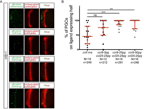
Different levels of chemokine receptor signaling leads to functionally same response. (A) Epifluorescence images of 10 hpf embryos. Embryos express the control RNA or the RNA encoding for Ccl25 in one half of the embryo and control RNA or different concentrations of RNA encoding for Ccr9 in PGCs. Merged images show the position of PGCs with respect to control or ligand-expressing domains (red). (B) Graphs show the quantitation of the migration of PGCs as the percentage of GFP-labeled cells located within the ligand-expressing domain of the embryos. 60 pg of mGFP-nanos was used to label PGCs in green and 40 pg of m-cherry mRNA was used for labeling the ligand expressing half of the embryo. For raw data see Figure 3—figure supplement 1—source data 1. |