
Fig. 1
- ID
- ZDB-FIG-180807-9
- Publication
- Lai et al., 2018 - Liver-directed microRNA-7a depletion induces nonalcoholic fatty liver disease by stabilizing YY1-mediated lipogenic pathways in zebrafish
- Other Figures
- All Figure Page
- Back to All Figure Page
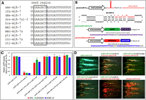
Design and validation of a miR-7a-sponge (miR7a-SP). (A) Alignment of the mature miR-7 sequence is perfectly conserved across many species, including honey bee (Apis mellifera; ame-miR-7), dog (Canis familiaris; cfa-miR-7), fruit fly (Drosophila melanogaster; dme-miR-7), zebrafish (Danio rerio; dre-miR-7a1-3), humans (Homo sapiens; hsa-miR-7), rhesus monkey (Macaca mulatta; mml-miR-7), mouse (Mus musculus; mmu-miR-7a), chimpanzee (Pan troglodytes; ptr-miR-7), rat (Rattus norvegicus; rno-miR-7) and frog (Xenopus tropicalis; xtr-miR-7). (B) Cloning of pri-miR-7a and miR7a-SP into b-Act expression vectors. Stem-loop structure of premiR-7a is shown, in which mature miR-7a is highlighted in red. (C) In vitro EGFP reporter assays were performed to confirm the direct interaction between miR-7a and the target sequences. ZFL, ZEM2S, and SOB-15 cells were transfected with pb-Act-eGFP-mir-7a-TS and pb-Act-eGFP-mir-7a or pb-Act-eGFP-mir-7a-SP plasmids, and the EGFP intensity was measured. **p < 0.01 and *p < 0.05. (D) In vivo EGFP reporter assays were performed to confirm the direct interaction between miR-7a and the target sequences in 6 dpf zebrafish larvae. |