
FIGURE
Fig. 4-S3
- ID
- ZDB-FIG-180801-26
- Publication
- LLeras Forero et al., 2018 - Segmentation of the zebrafish axial skeleton relies on notochord sheath cells and not on the segmentation clock
- Other Figures
- All Figure Page
- Back to All Figure Page
Fig. 4-S3
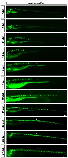
her1−/−;her7−/−;entpd5:kaede larvae develop axial segmentation in a disorganized manner, occasionally missing one or two segments or inserting additional segments. Fusions of adjacent chordacentra, non-regular entpd5+ segment shapes, thicker and thinner segments and a transient bending of the axis (asterisk) can be seen. Virtual time lapse of one larva from 7 dpf to 27 dpf. Scale bars are 300 µm. |
Expression Data
Expression Detail
Antibody Labeling
Phenotype Data
Phenotype Detail
Acknowledgments
This image is the copyrighted work of the attributed author or publisher, and
ZFIN has permission only to display this image to its users.
Additional permissions should be obtained from the applicable author or publisher of the image.
Full text @ Elife