
Fig. 3-S1
- ID
- ZDB-FIG-180724-6
- Publication
- Janjuha et al., 2018 - Age-related islet inflammation marks the proliferative decline of pancreatic beta-cells in zebrafish
- Other Figures
- All Figure Page
- Back to All Figure Page
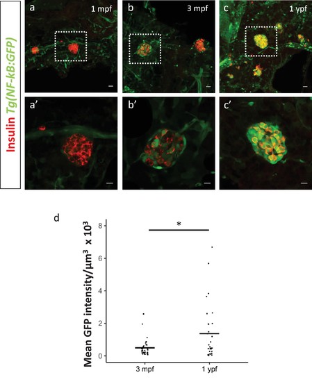
Activation of NF-kB signaling in beta-cells of the secondary islets with age. (a,b,c) Confocal stack of secondary islets from Tg(NF-kB:GFP) animals at 1 mpf, 3 mpf and 1 ypf. Beta-cells were labeled using an insulin antibody (red). NF-kB:GFP reporter expression is shown in green. Scale bars 20 µm. (a’,b’,c’) Insets show high-magnification single planes of the confocal stacks corresponding to the regions outlined using white dotted-lines in the top panels. Scale bar 10 µm. (d) Graph showing the total normalized GFP fluorescence intensity of the secondary islets from 3 mpf (n = 9 fish, secondary islets = 32) and 1 ypf (n = 8, secondary islets = 30) animals. Each dot represents one islet (two-tailed t-test, *p<0.05). |