
Fig. 6
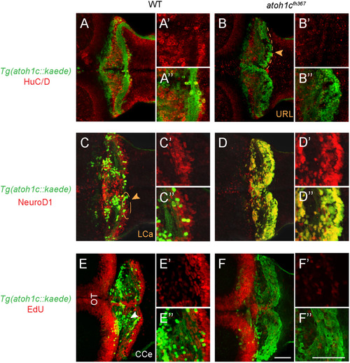
atoh1cfh367 cerebellar cells accumulate as post-mitotic but undifferentiated granule cell progenitors. A,B: The majority of Tg(atoh1c::kaede)+ cells (green) in the atoh1c mutant are located within the URL and do not express HuC/D (red) at 5 dpf (B) indicating that they are not post-mitotic neurons. C,D: atoh1cfh367 cells (green) in the URL are positive for NeuroD1 (red, D). E,F: atoh1cfh367 cells (green) in the URL do not incorporate EdU (red, F). Strong EdU incorporation anterior to the cerebellum in both wild-type and mutant is in the optic tectum (OT). Dorsal views with anterior to the left. Scale bars: 50 μM. |
| Gene: | |
|---|---|
| Antibodies: | |
| Fish: | |
| Anatomical Terms: | |
| Stage: | Day 5 |
| Fish: | |
|---|---|
| Observed In: | |
| Stage: | Day 5 |
Reprinted from Developmental Biology, 438(1), Kidwell, C.U., Su, C.Y., Hibi, M., Moens, C.B., Multiple zebrafish atoh1 genes specify a diversity of neuronal types in the zebrafish cerebellum, 44-56, Copyright (2018) with permission from Elsevier. Full text @ Dev. Biol.