
Fig. 2
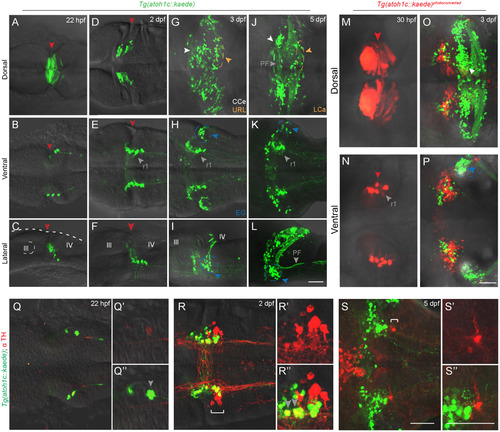
atoh1c-expressing progenitors give rise to to ventral r1 and cerebellar granule neurons. A-L: Tg(atoh1c::kaede) transgene expression in fixed embryos stained with anti-Kaede antibody in dorsal (A,D,G,J), ventral (B,E,H,K) and lateral (C,F,I,L) views at 22 hpf (A-C), 2 dpf (D-F), 3 dpf (G-I) and 5 dpf (J-L). Arrowheads follow the color code described in Fig. 1 legend or are labeled as follows: PF: parallel fibers, r1: MHB-derived neurons in ventral r1. M-P: live imaging after photoconversion of Kaede (green to red) of MHB atoh1c+ cells at 22 hpf confirms that this Tg(atoh1c::kaede)+ progenitor domain gives rise to ventral r1 neurons. Dorsal (M-O) and ventral focal planes (N-P). Q-S: Tg(atoh1c::kaede)+ cells (green) transiently express TH (red; gray arrowheads) from 22 hpf (Q) to 2 dpf (R) and lie adjacent to the LC (indicated by white bracket). All images oriented with anterior to the left at time points as indicated. Scale bars: 50 μM. |
| Genes: | |
|---|---|
| Antibody: | |
| Fish: | |
| Anatomical Terms: | |
| Stage Range: | 26+ somites to Day 5 |
Reprinted from Developmental Biology, 438(1), Kidwell, C.U., Su, C.Y., Hibi, M., Moens, C.B., Multiple zebrafish atoh1 genes specify a diversity of neuronal types in the zebrafish cerebellum, 44-56, Copyright (2018) with permission from Elsevier. Full text @ Dev. Biol.