
Fig. 3
- ID
- ZDB-FIG-180703-9
- Publication
- Takaki et al., 2018 - A zebrafish model for ocular tuberculosis
- Other Figures
- All Figure Page
- Back to All Figure Page
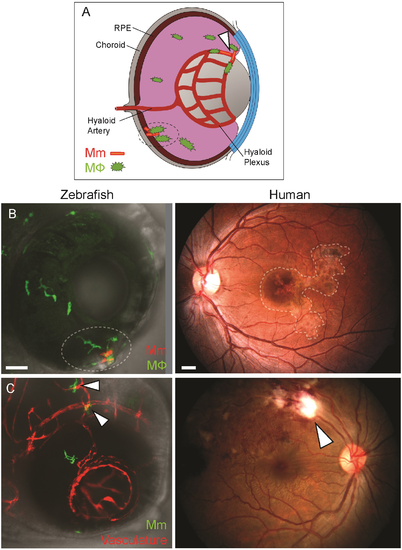
Anatomical localization of intraocular granuloma after M. marinum infection. (A) Schematic representation showing localization of granulomas near the retinal vasculature (arrowhead), and in retinal pigment epithelium-choroid complex (dotted circle). (B) Localization of intraocular granulomas (within dotted regions); in the outer eye of zebrafish larvae corresponding to the retinal pigment epithelium-choroid complex, and in the choroid in human ocular TB. (C) Localization of perivascular infection (arrowheads); as seen as bacterial aggregates in close association of blood vessels in zebrafish, and retinal periphlebitis associated with focal chorioretinitis overlying the blood vessel in human ocular TB. (B-C) Confocal images of ocular infection in Tg(mpeg:YFP) and Tg(kdrl:dsRed2) zebrafish, and fundus photographs of human ocular TB. Scale bars, 25 μm and 750 μm, respectively. |
| Fish: | |
|---|---|
| Condition: | |
| Observed In: | |
| Stage: | Adult |