
FIGURE
Fig. S4
- ID
- ZDB-FIG-180702-5
- Publication
- Hocking et al., 2018 - Morphogenetic defects underlie Superior Coloboma, a newly identified closure disorder of the dorsal eye
- Other Figures
- All Figure Page
- Back to All Figure Page
Fig. S4
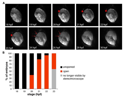
Dynamics of the zebrafish superior ocular sulcus. (A) Time-lapse images showing lateral views of the eye of a Tg(rx3:GFP) embryo. The superior ocular sulcus appears as a narrow groove across the dorsal retina at ~20 hpf (red asterisk), becomes wider by 24 hpf (red arrows) and disappears after 26 hpf. (B) Timing of SOS as viewed under a stereomicroscope. The wide and shallow phase is not visible by stereomicroscope, so the red bars indicate the percentage of embryos with a narrow and distinct sulcus. |
Expression Data
| Gene: | |
|---|---|
| Fish: | |
| Anatomical Terms: | |
| Stage Range: | 14-19 somites to Prim-15 |
Expression Detail
Antibody Labeling
Phenotype Data
Phenotype Detail
Acknowledgments
This image is the copyrighted work of the attributed author or publisher, and
ZFIN has permission only to display this image to its users.
Additional permissions should be obtained from the applicable author or publisher of the image.
Full text @ PLoS Genet.