
Fig. 2
- ID
- ZDB-FIG-180702-1
- Publication
- Hocking et al., 2018 - Morphogenetic defects underlie Superior Coloboma, a newly identified closure disorder of the dorsal eye
- Other Figures
- All Figure Page
- Back to All Figure Page
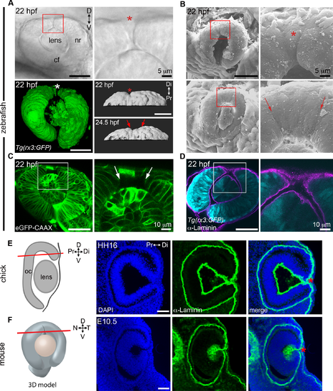
The superior ocular sulcus in zebrafish, chick and mouse. (A) Zebrafish eyes displaying superior ocular sulci (SOS) marked by an asterisk or arrows. Top row: lateral view DIC image of the eye of a live embryo, photographed on a compound microscope. Enlarged view is shown in panel on right. Bottom row: Left, lateral view surface projection of the eye of a live Tg(rx3:GFP) embryo; Right, surface projection dorsal views of eyes from a Tg(rx3:GFP) embryo. (B) Scanning electron micrographs showing SOS at narrow (top row) and wide (bottom row) phases. Red boxes denote regions enlarged in panels on the right. (C) Single optical section, lateral view, through the eye of an embryo injected with eGFP-CAAX mRNA to label the cell membranes, with right panel showing enlarged view of boxed area. (D) Single optical section, lateral view, through eye of Tg(rx3:GFP) embryo (cyan) immunolabelled for Laminin to highlight the basal lamina (magenta). (E) Diagram showing chick eye with red line demonstrating the plane of section employed on the right. Representative horizontal section through the dorsal eye of a HH16 chick, stained with a Laminin antibody (green) and DAPI (blue). A dorsal, Laminin-lined space is evident in the distal portion of optic cup (asterisk). (F) Diagram showing 3D model of an embryonic eye with red line demonstrating plane of section for both mouse and chick sections. Right three panels are a representative horizontal section through the dorsal eye of an embryonic day 10.5 (E10.5) mouse, stained with a Laminin antibody (green) and DAPI (blue). A dorsal, Laminin-lined space is evident in the distal portion of optic cup (asterisk). Except where noted, scale bars are 50 μm. cf, choroid fissure; D-V, dorsal-ventral; HH, Hamburger Hamilton embryonic stage; hpf, hours post fertilization; N-T, nasal-temporal; nr, neural retina; Pr-Di, proximal-distal. |
| Gene: | |
|---|---|
| Fish: | |
| Anatomical Terms: | |
| Stage: | 26+ somites |