Fig. S6
|
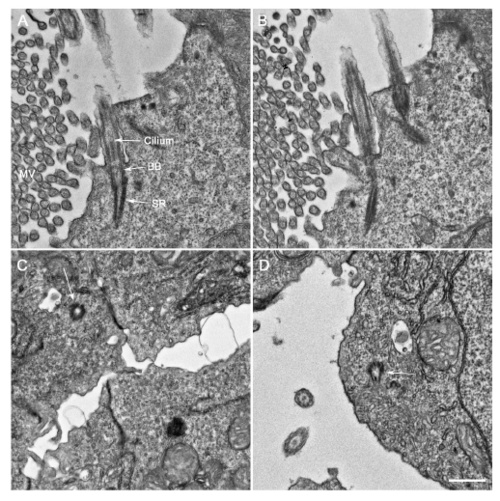
Undocked centrioles/basal bodies in 6 dpf pam-/- embryos Transverse sections through wildtype and pam-/- embryos (6 dpf). (A, B) Adjacent sections through a cell lining the pronephros of a 6 dpf wildtype embryo showing two basal bodies (BB) with striated rootlets (SR) docked at the plasma membrane with attached membrane-bound ciliary axonemes (cilium) extending into the pronephric lumen; numerous microvilli (MV) are evident. (C, D) Undocked centrioles/basal bodies (arrows) located within the cytoplasm of cells lining the pronephros of a 6 dpf pam-/- embryo and oriented approximately parallel to the pronephros long axis as are the cytosolic axonemes (see Fig. 5H); few cilia are present in the lumen and microvilli are absent. Bar = 500 nm. |
| Fish: | |
|---|---|
| Observed In: | |
| Stage: | Day 6 |

