
Fig. 5
- ID
- ZDB-FIG-180627-38
- Publication
- Sedykh et al., 2017 - Zebrafish Rfx4 controls dorsal and ventral midline formation in the neural tube
- Other Figures
- All Figure Page
- Back to All Figure Page
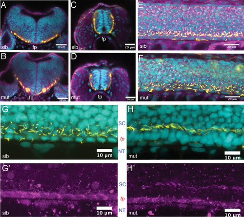
Ciliogenesis is not overtly affected in the spinal cord of rfx4 mutant embryos. A–F: 24‐hpf embryos derived from an rfx4uw8013/+ incross were immunostained for acetylated tubulin (yellow) and gamma‐tubulin (purple) to visualize ciliary axonemes and basal bodies, respectively. Nuclei were counterstained with DAPI (cyan). Confocal cross‐sections through the hindbrain (A,B) and spinal cord (C,D). E: confocal sagittal z‐stack through the spinal cord of a heterozygous sibling shows normal distribution of primary (short) and motile (long) cilia. F: Equivalent z‐stack through a homozygous mutant sibling shows presence of both primary and motile cilia. G,H′: rfx4uw8013/+ incross progeny were immunostained for acetylated tubulin (yellow in G,H) and a motile cilia component rsph9 (purple; G′, H′) to visualize ciliary axonemes of primary vs motile cilia. Nuclei were counterstained with DAPI (cyan). G: confocal sagittal z‐stack through the ventral portion of the spinal cord of a heterozygous sibling shows normal medial floor plate morphology and normal distribution of motile (long) cilia produced by both medial and lateral floorplate. H: Equivalent z‐stack through a homozygous mutant sibling shows presence of long cilia in the absence of identifiable floor plate. fp, floor plate; SC, spinal cord; NT, notochord. Scale bar = 30 μm in A,B; 20 μm in C–F; 10 μm in G,H. |
| Fish: | |
|---|---|
| Observed In: | |
| Stage: | Prim-5 |