Fig. 4
- ID
- ZDB-FIG-180626-22
- Publication
- Takei et al., 2018 - High-Sensitivity and High-Resolution In Situ Hybridization of Coding and Long Non-coding RNAs in Vertebrate Ovaries and Testes
- Other Figures
- All Figure Page
- Back to All Figure Page
|
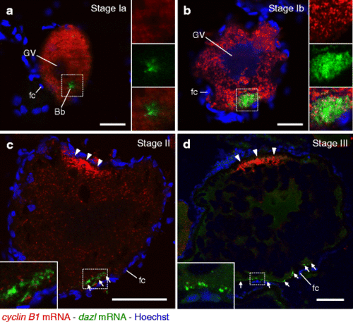
Double fluorescence in situ hybridization of cyclin B1 (red) and dazl (green) mRNAs in zebrafish ovaries. DNA is shown in blue. a A follicle consisting of stage Ia oocyte. Insets are enlarged views of the boxed region showing cyclin B1 mRNA (upper), dazl mRNA (middle) and a merged image (lower). b A follicle consisting of stage Ib oocyte. Insets are enlarged views of the boxed region showing cyclin B1 mRNA (upper), dazl mRNA (middle) and a merged image (lower). c A follicle consisting of stage II oocyte. The inset is an enlarged view of the boxed region. d A follicle consisting of stage III oocyte. The inset is an enlarged view of the boxed region. Arrowheads indicate cyclin B1 RNA granules localized at the animal polar cytoplasm of oocytes. Arrows indicate dazl RNA granules distributed in the vegetal polar cytoplasm of oocytes. GV, germinal vesicle; Bb, Balbiani body; fc, follicle cells. Bars: 20 μm in a and b, 50 μm in c and d |
| Genes: | |
|---|---|
| Fish: | |
| Anatomical Terms: | |
| Stage: | Adult |

