
FIGURE
Fig. 3
- ID
- ZDB-FIG-180615-11
- Publication
- Brown et al., 2017 - Neuregulin-1 is essential for nerve plexus formation during cardiac maturation
- Other Figures
- All Figure Page
- Back to All Figure Page
Fig. 3
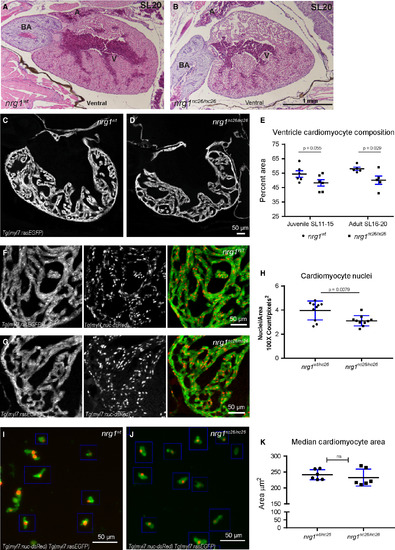
Reduced trabecular myocardium in nrg1nc26 mutants due to lower cellularity. (A) Representative cross section of the heart in H&E‐stained section of formaldehyde‐fixed, paraffin‐embedded nrg1WT and nrg1nc26/nc26 adult fish at SL = 20, N = 3. BA = Bulbous Arteriosus, V = Ventricle, A = Atrium. Scale bar 100 μm. (C–E) Intracardiac density measured from confocal 9.6 μm cross section of hearts from Tg(myl7:rasGFP) fish. (C, D) Representative images, SL = 13. (E) Intracardiac myocardial density quantified as the per cent area occupied by rasGFP+ cells in the outer curvature relative. (F–H) Cardiomyocyte numbers measured from confocal 9.6 μm cross section of hearts from double transgenic Tg(myl7:rasGFP);Tg(myl7:nuc‐dsRED) fish (F, G) Representative images, SL = 13. (H) Relative number of cardiomyocytes measured as from cross sections as the ratio of dsRED‐positive nuclei to rasGFP‐positive pixels in fish SL11‐SL20. (I–K) Cardiomyocyte size measured in mononuclear cells dissociated from Tg(myl7:rasGFP);Tg(myl7:nuc‐dsRED) hearts. (I, J) Representative dissociated cell preparations. (K) Median area of N > 200 cells per sample with 1–2 hearts per sample, N = 6 biological replicates.
|
Expression Data
Expression Detail
Antibody Labeling
Phenotype Data
| Fish: | |
|---|---|
| Observed In: | |
| Stage: | Adult |
Phenotype Detail
Acknowledgments
This image is the copyrighted work of the attributed author or publisher, and
ZFIN has permission only to display this image to its users.
Additional permissions should be obtained from the applicable author or publisher of the image.
Full text @ J. Cell. Mol. Med.