
FIGURE
Fig. S36
- ID
- ZDB-FIG-180611-55
- Publication
- Triemer et al., 2018 - Superresolution imaging of individual replication forks reveals unexpected prodrug resistance mechanism
- Other Figures
- All Figure Page
- Back to All Figure Page
Fig. S36
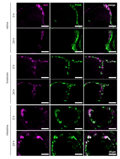
Staining of AzC and PCNA in the retina, forebrain and intestine after termination of AzC treatment. Three day old zebrafish were incubated in 5 mM AzC, followed by a 24 h recovery period in the presence of 1 mM F-ara-EdU. Proliferating cells were visualized with PCNA immunolabeling. AzC was stained using SiR alkyne, Cu(I), and THPTA. |
Expression Data
Expression Detail
Antibody Labeling
Phenotype Data
Phenotype Detail
Acknowledgments
This image is the copyrighted work of the attributed author or publisher, and
ZFIN has permission only to display this image to its users.
Additional permissions should be obtained from the applicable author or publisher of the image.
Full text @ Proc. Natl. Acad. Sci. USA