
- Title
-
Superresolution imaging of individual replication forks reveals unexpected prodrug resistance mechanism
- Authors
- Triemer, T., Messikommer, A., Glasauer, S.M.K., Alzeer, J., Paulisch, M.H., Luedtke, N.W.
- Source
- Full text @ Proc. Natl. Acad. Sci. USA

Metabolic incorporation of AzC and reversible inhibition of DNA synthesis in the retinal stem cell niche of zebrafish larvae. (A) Staining patterns of AzC and AmdU after 24-h treatment of 3-d-old fish with 10 mM AmdU or 5 mM AzC, fixation, and staining with SiR alkyne. (B) Lack of overlap between nucleolin immunofluorescent staining (arrows) and AzC after 24-h treatment of 3-d-old zebrafish with 5 mM AzC. (C) Overlap between AzC and PCNA staining after 24-h treatment of 3-d-old zebrafish with 5 mM AzC. (D) Inhibition of DNA synthesis by addition of AzC or ara-C. Three-day-old fish were incubated with 5 mM AzC, ara-C, or AmdU for 3 h, followed by coincubation with 1 mM F-ara-EdU for 21 h. F-ara-EdU was visualized using Alexa Fluor 488 azide. (E) Resumption of DNA synthesis after removal of AzC or ara-C. Following a 24-h pulse of 5 mM AzC, ara-C, or AmdU, the fish were moved into fresh water containing 1 mM F-ara-EdU for a 60-h recovery period before fixation and staining. (F) Movement of AzC-labeled DNA out of the retinal stem cell niche according to an AzC pulse, F-ara-EdU chase experiment as described in E. See SI Appendix, Figs. S29–S37 for the corresponding images of the liver, intestine, and brain. |
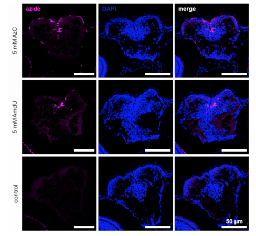
AzC and AmdU incorporation and staining in proliferating cells of the zebrafish forebrain. Three day old zebrafish were incubated in 5 mM AzC or AmdU for 24 h, and AzC and AmdU were stained using SiR alkyne, Cu(I), and THPTA. The control animals were treated the same but did not receive any azido nucleoside analog. |

AzC incorporation and staining in (A) the retina, (B) forebrain, and (C) the intestine and liver. Three day old zebrafish were incubated in 10 mM AzC for 24 h, and AzC was labeled using SiR alkyne, Cu(I), and THPTA. The control animals were treated the same but did not receive any azido nucleoside analog. Staining was detected in all other proliferating areas throughout the body. |
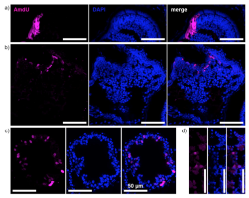
AmdU incorporation and staining in (A) the retina, (B) forebrain, (C) the intestine, and (D) liver. Three day old zebrafish were incubated in 10 mM AzC for 24 h, and AzC was labeled using SiR alkyne, Cu(I), and THPTA. The control animals were treated the same but did not receive any azido nucleoside analog. Staining was detected in all other proliferating areas throughout the body. |
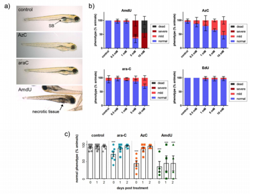
Toxicity of nucleoside analogs in zebrafish larvae. (A) Representative photographs of 96 h old zebrafish larvae following 24 h incubations with 5 mM of each nucleoside analog. (B) Summary of data for 72 h old zebrafish larvae incubated with various concentrations (0.5 – 10 mM) of each nucleoside for 24 h and analyzed for phenotypic changes. Fish treated with AzC or ara-C lacked swim bladder inflation (corresponds to “mild” phenotype) whereas fish treated with AmdU displayed necrotic tissue and horizontal body positioning (corresponds to severe phenotype). (C) 72 h old zebrafish larvae were incubated with 10 mM of nucleoside analog for 24h, washed, and placed into media lacking nucleoside analog for up to 2 days (recovery). After a recovery period of 1 – 2 days, fish treated with ara-C and AzC regained a normal morphology, whereas AmdU-treated fish showed no signs of recovery. All experiments were repeated at least 3 times. Data are presented as mean ± SEM, *** p < 0.001 relative to controls; SB, swim bladder. |
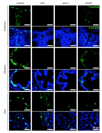
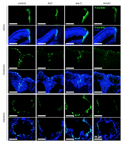
Inhibition of DNA synthesis by AzC and ara-C in stem cell niches of developing zebrafish. Three day old zebrafish were incubated in 5 mM AzC, ara-C or AmdU for 3 h, followed by co-incubation with 1 mM F-ara-EdU for 21 h. F-ara-EdU was stained using Alexa 488 azide, Cu(I), and THPTA. |
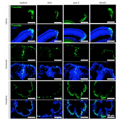
Resumption of DNA synthesis in retina, forebrain and intestine after termination of AzC or ara-C treatment. Three day old zebrafish were incubated in 5 mM AzC, ara-C or AmdU for 24 h, followed by a 24 h recovery period in the presence of 1 mM F-araEdU. F-ara-EdU was visualized using Alexa 488 azide, Cu(I), and THPTA. |
Resumption of DNA synthesis in retina, forebrain and intestine after termination of AzC or ara-C treatment. Three day old zebrafish were incubated in 5 mM AzC, ara-C or AmdU for 24 h, followed by a 60 h recovery period in the presence of 1 mM F-araEdU. F-ara-EdU was visualized using Alexa 488 azide, Cu(I), and THPTA. |
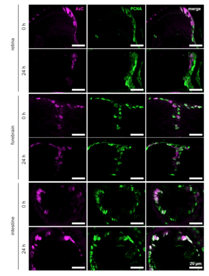
Staining of AzC and PCNA in the retina, forebrain and intestine after termination of AzC treatment. Three day old zebrafish were incubated in 5 mM AzC, followed by a 24 h recovery period in the presence of 1 mM F-ara-EdU. Proliferating cells were visualized with PCNA immunolabeling. AzC was stained using SiR alkyne, Cu(I), and THPTA. |

Staining of AzC and PCNA in the retina, forebrain and intestine after termination of AzC treatment. Three day old zebrafish were incubated in 5 mM AzC, followed by a 60 h recovery period in the presence of 1 mM F-ara-EdU. Proliferating cells were visualized with PCNA immunolabeling. AzC was stained using SiR alkyne, Cu(I), and THPTA. |