
Fig. 4
- ID
- ZDB-FIG-180523-16
- Publication
- Shin et al., 2017 - Saprolegnia parasitica Isolated from Rainbow Trout in Korea: Characterization, Anti-Saprolegnia Activity and Host Pathogen Interaction in Zebrafish Disease Model.
- Other Figures
- All Figure Page
- Back to All Figure Page
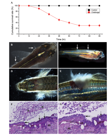
Survival rate, gross and histopathological observation of zebrafish challenged with Saprolegnia parasitica. A, Cumulative survival percentage of juvenile zebrafish challenged with S. parasitica; B, C, Typical whitish mycelium of S. parasitica near to gill and trunk (B) and methylene blue stained S. parasitica mycelium (C) in all over the trunk of infected juvenile zebrafish; D, E, S. parasitica infection near to the gills (D) and trunk (E) of adult zebrafish; F, G, Histopathological observations of Saprolegnia infected skin tissue. Damage on the epithelium (F and G; black arrow), tissues near to scales (F and G; white arrows) and muscle (F and G; m). S. parasitica hyphae (F and G; h) grown on the epithelial tissue. Over-activation of epithelial goblet cells (F and G; black dotted arrows). Large number of immune cells (F and G; i) infiltration near to infection area (×200); G, Magnified image (×400) of Fig. 4F (scale bars: F = 100 µm, G = 50 µm). Stained with Periodic acid Schiff. |