
Fig. 3
- ID
- ZDB-FIG-180523-15
- Publication
- Shin et al., 2017 - Saprolegnia parasitica Isolated from Rainbow Trout in Korea: Characterization, Anti-Saprolegnia Activity and Host Pathogen Interaction in Zebrafish Disease Model.
- Other Figures
- All Figure Page
- Back to All Figure Page
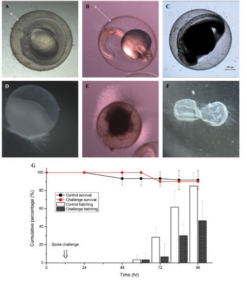
Morphology, survival rate, and hatching delay percentage of Saprolegnia parasitica challenged zebrafish embryos. A, B. S. parasitica zoospores were germinated and started growing on the surface of chorion at 3-hr post infection (hpi) and 6 hpi. White dotted line indicates the grown length of mycelium; C, S. parasitica mycelium penetrated into a whole embryo and grown inside the embryo at 30 hpi; D, E, Abundant mycelium growth in the dead embryos at 3 hpi and 12 hpi; F, Mycelium growth on the chorion which remained after hatching; G, Cumulative survival rate and hatching delay percentage of embryo exposed to S. parasitica secondary zoospores. Survival and hatching rate percentage of embryos were the means of 6 individual groups (10 embryos in each group). |