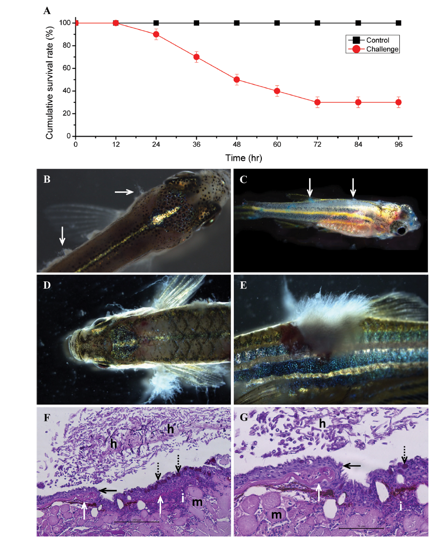
Fig. 4 Survival rate, gross and histopathological observation of zebrafish challenged with Saprolegnia parasitica. A, Cumulative survival percentage of juvenile zebrafish challenged with S. parasitica; B, C, Typical whitish mycelium of S. parasitica near to gill and trunk (B) and methylene blue stained S. parasitica mycelium (C) in all over the trunk of infected juvenile zebrafish; D, E, S. parasitica infection near to the gills (D) and trunk (E) of adult zebrafish; F, G, Histopathological observations of Saprolegnia infected skin tissue. Damage on the epithelium (F and G; black arrow), tissues near to scales (F and G; white arrows) and muscle (F and G; m). S. parasitica hyphae (F and G; h) grown on the epithelial tissue. Over-activation of epithelial goblet cells (F and G; black dotted arrows). Large number of immune cells (F and G; i) infiltration near to infection area (×200); G, Magnified image (×400) of Fig. 4F (scale bars: F = 100 µm, G = 50 µm). Stained with Periodic acid Schiff.
Image
Figure Caption
Acknowledgments
This image is the copyrighted work of the attributed author or publisher, and
ZFIN has permission only to display this image to its users.
Additional permissions should be obtained from the applicable author or publisher of the image.
Full text @ Mycobiology

